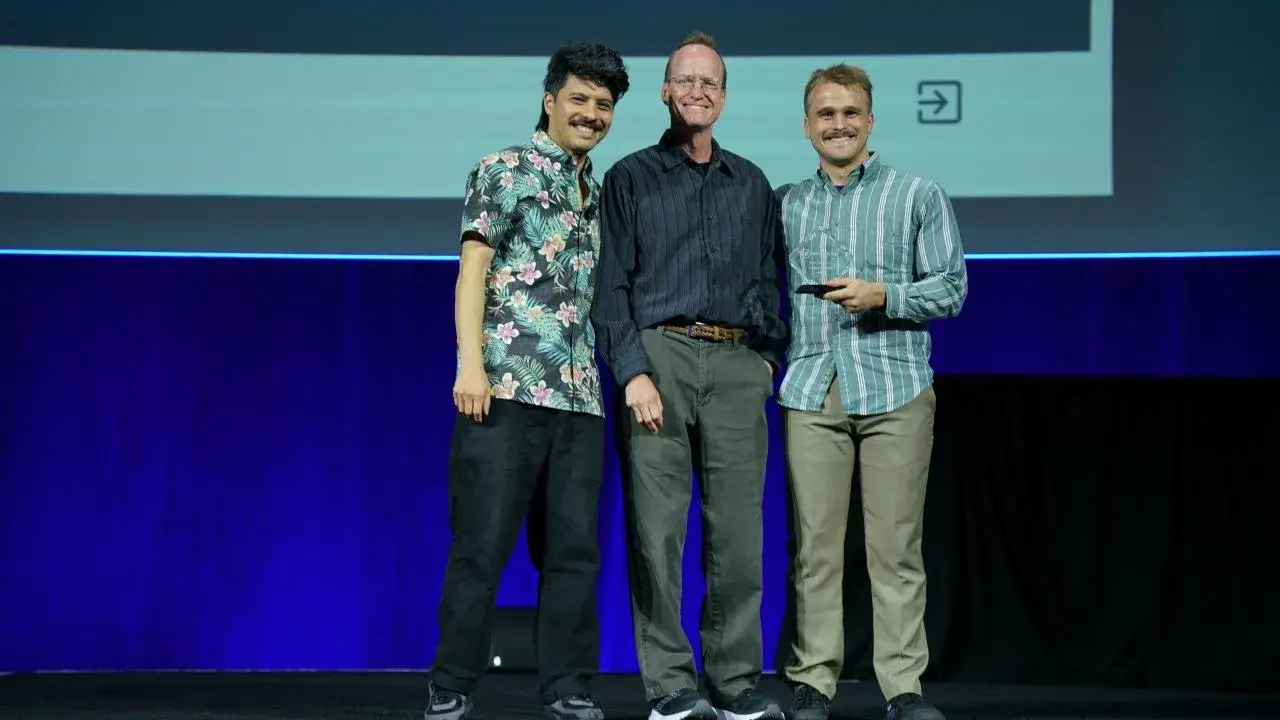
At SIGGRAPH, Jack Myers cuts into a virtual body, opens the incision and removes a structure without waiting for a heavy solver to catch up. The demonstration won the 2026 Real-Time Live! Audience Choice award,13 yet the more revealing part sits in the paper by Myers and Tim McGraw: the displayed mechanics are not meant to reproduce the mechanics of human tissue faithfully.2
The authors call their method non-physical animation. That would be a serious weakness in a surgical simulator, but their target is different: let somebody explore anatomy by cutting, tearing and removing pieces while the model continues to respond at interactive speed.2
Fake tissue
An incision makes a digital body much harder to manage. Before the blade passes, two regions belong to the same volume; afterwards new surfaces exist, both sides need to separate, and a detached piece may have to move independently, all while the software keeps drawing the next frame. Adding faithful material mechanics for every tissue makes that loop considerably more expensive.
McGraw and Myers instead use position-based dynamics. Particles sit on a grid and belong to overlapping regions whose shape constraints pull the model toward coherent configurations; for this application, the important property of those constraints is that they are breakable.2
When a cut crosses a region, the affected constraint splits, so particles on opposite sides no longer have to behave as one piece; repeated throughout the volume, that mechanism allows an incision to open or a part to separate without solving the real mechanics of flesh every frame.2
The paper is unusually explicit about the consequence. Parameters controlling apparent stiffness have no direct mapping to Young's modulus or other measured material properties.2 The anatomy is documented; the “material” holding it together is invented for interaction.
Real slices
The visual source makes that separation stranger still. Purdue reuses the U.S. National Library of Medicine's Visible Human Project, an archive assembled in the 1990s from two cadavers using colour cryosection photography alongside CT and MRI.4
Anatomical photographs of the male body were taken at one-millimetre intervals, while the female dataset reduced the spacing to 0.33 mm and contains more than 5,000 anatomical images.4 Three decades later, those photographs still provide far more visual detail than the mechanical grid that moves them.
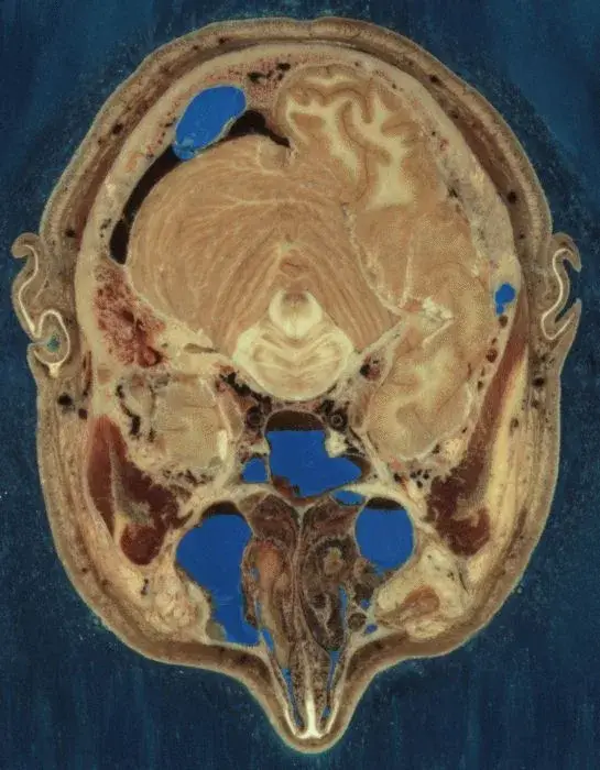
Inside Dissectible Anatomy, volumetric splats carry colour and detail sampled from those cryosections, whereas a lower-density particle grid controls deformation.2 The resolutions deliberately diverge, allowing relatively few mechanical degrees of freedom to move a visually rich volume.
That split explains much of the performance and also gives old scientific data a new role. The photographs provide internal content; the dynamic layer turns them from a stack of slices into something a user is allowed to damage.
Cut fast
Repeated cuts are awkward for an ordinary triangle mesh, because every fracture creates topology that has to remain valid while exposing a new surface, and the problem gets worse when a user immediately pulls on what was just cut.
The Purdue system uses a signed distance function sampled at its splats. Cutting and tearing update that representation together with the mechanical constraints rather than rebuilding a clean mesh after each gesture,2 and the paper formalises three anatomy-relevant fracture operations: cutting, tearing and excision.
Where anatomical segmentation is available, the software also associates names and boundaries with structures. Purdue demonstrates an organ being separated from the body, after which fresh constraints keep the excised piece usable as an object in its own right.1
GPU compute shaders carry the implementation. Published measurements were taken on a PC with an NVIDIA GeForce RTX 4070, and for the tested workloads the authors report faster performance than their MPM and FEM comparisons.2 The useful result is less a spectacular polygon count than the ability to keep responding after the model stops having fixed topology.
The cost
A mechanical grid coarser than the visible data eventually leaves traces. The authors report possible artefacts around hard/soft tissue boundaries because tissue class belongs to the splats while mechanical behaviour is averaged onto particles;2 an unsegmented dataset loses more information still, receiving homogeneous apparent properties instead of distinct behaviour for different tissues.
Rendering also grows harder as dissection exposes the inside of the model. More internal surface means more splats inside the visible narrow band, and the paper lists that increase in rendering cost among the problems left for future optimisation.2
The more subtle problem never appears in a benchmark. Motion that looks convincing can imply more mechanical truth than the model contains, especially when its colour comes from actual anatomical sections, so an educational interface has to keep two kinds of fidelity separate: location, shape, colour and sometimes identity are strongly grounded in the source data, while mechanical response is knowingly approximate.
Not surgery
That distinction rules out treating the demo as training for an operative gesture. McGraw and Myers note that a surgical application would require the mechanical accuracy they deliberately give up; their aim here is plausible animation fast enough for exploration.2
The choice is less strange than it sounds. An anatomy student may not need a correct prediction of cutting force to learn that one structure passes behind another, where an organ sits, or what becomes visible after opening a layer; the approximation matters much more once the lesson depends on transferring resistance, touch or a clinical movement into the physical world.
The SIGGRAPH presentation works because the first use is immediately legible: cut, open, inspect. It never measures the second.
Missing test
The team has not yet shown that students learn more effectively with the system. Its paper instead lists a testbed for user studies of anatomy learning outcomes as future work,2 while Purdue describes the tool as a safe and accessible way to practise and review and notes that the researchers do not expect it to replace physical dissection entirely.1
Audience Choice at SIGGRAPH establishes something narrower: the interaction survives a live presentation and communicates itself within seconds. Moving from an excellent graphics demonstration to an effective teaching tool requires the result that has not been published yet, namely what a student remembers after closing the virtual body.
The next interesting version may therefore look almost identical. If deliberately approximate mechanics produce a better mental model of anatomy, Purdue will have shown that in educational simulation the most useful fidelity is not necessarily the one that is easiest to see.